
For more than a year, Rhys Hibbert saw his field of vision narrow. He stumbled and had to walk with sticks. The cause: a tumor measuring only 11 millimeters, located in the pituitary gland, in the immediate vicinity of the optic nerves.
In May 2026, this 48-year-old Briton became the first patient in the world to undergo a neurosurgical procedure during which artificial intelligence analyzed the images of the operation in real time. Its role: to help surgeons identify the structures to be preserved to remove the tumor without damaging the optic nerves.
When you wake up, the change is immediate. “When I woke up…I could clearly see everything in the room“, he says. A week later, he was walking without glasses or poles. “I feel like I have a 360 degree panoramic view of everything around me“.
It all starts with feeling unwell during a walk
In December 2024, Rhys was taking his daily walk when he suddenly felt unwell before losing consciousness and having a seizure.
“My tumor was discovered unexpectedly in December 2024. I was taking my usual daily walk when I suddenly felt unwell and lost consciousness.”he says. After hospital examinations, doctors discovered a tumor measuring around 11 millimeters in his pituitary gland.
Its symptoms will then worsen: significant hormonal imbalances and vision problems. “I started using walking poles after tripping several times because I couldn’t see the lower part of my vision.”he explains.
Surgery becomes necessary. But the location of the brain tumor makes the intervention particularly delicate.
In this area, a millimeter can change everything
The size of a marble, the pituitary gland is located at the base of the brain, surrounded in particular by the optic nerves and large blood vessels. In this extremely narrow space, a deviation of even a millimeter can lead to blindness, stroke or, in the most severe cases, death.
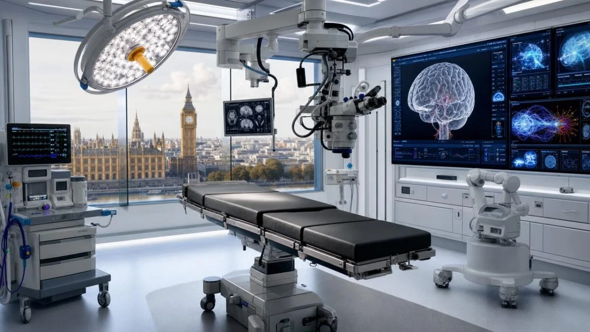
This is where artificial intelligence comes in. During the operation, an endoscopic camera films the operated area. Unlike systems which only analyze scans taken before the intervention, the tool developed by University College London analyzes live surgical video.
It helps identify critical anatomical structures, including nerves and vessels, but also instruments and their interactions with tissues. The objective is to allow the surgeon to better identify areas at risk while removing as much tumor as possible under the best possible safety conditions.
But the AI does not make any decisions for the doctor. The procedure remains entirely under the control of the neurosurgeon, Professor Hani Marcus, assisted by Danyal Khan.
For Rhys, the interest is obvious: “From a patient safety perspective, I absolutely see the benefit. There are no signposts inside our heads.”.
An AI trained on hundreds of operations
The system was developed by the UCL Hawkes Institute and trained on hundreds of carefully annotated pituitary surgery videos. Researchers taught him to recognize different anatomical structures, surgical instruments and interactions between tissues.
“By learning from hundreds of surgical videos, he was exposed to a diversity of surgical examples that would take a surgeon many years to encounter.”explains Dr. Sophia Bano, technical manager of the system. It is designed to “help recognize critical anatomy, surgical instruments and tissue interactions in real time”.
The technology had already been evaluated as a training tool for surgeons. This intervention marks a new stage: for the first time, it provided assistance
during a real operation.
And for Rhys, the result is particularly concrete. “There is a risk that a patient will come out of surgery with worse vision. But I think my vision is a testament to their precision.”he believes.
Rhys agreed to become the first for those who come after
Rhys could have refused to participate in this world premiere. He chose to accept, convinced that medical research also relies on patient engagement.
“If patients are not willing to participate in research, how can doctors learn and how can medicine advance?he asks.
His choice takes on particular resonance in this hospital founded in 1859, presented by the establishment as the first in the world dedicated to neurosurgery.
“The National Hospital for Neurology and Neurosurgery was founded in 1859… and was the first hospital in the world devoted exclusively to neurosurgery. So, to me, it seems very fitting that the same hospital is also the first in the world to perform AI-assisted neurosurgery.”he emphasizes.
Before his operation, he even brought two boxes of chocolates to the operating room to thank the team. “It’s not thanks to me. It’s thanks to the team of surgeons, the anesthetists and, in fact, the entire support team… who make this kind of success possible.”he insists.
A world first, but not yet a revolution
This operation does not mean that AI will replace neurosurgeons. The system remains an assistance tool, used as part of a clinical trial. Its effectiveness and interest will still need to be evaluated in more patients.
The trial is funded in particular by the National Institute for Health and Care Research and Google, with the support of several British research and surgical organizations.
For James Frith, British Minister for Health Innovation, safeguards remain essential: “AI must have appropriate safeguards and we will always ensure that security is taken seriously.”.
The interest of this first intervention is therefore less to dedicate “AI surgery” than to show what assistance capable of instantly analyzing the images of an operation could bring to surgeons.
In a region of the brain where a few millimeters can separate a successful intervention from irreversible loss of sight, this help could, tomorrow, count.