
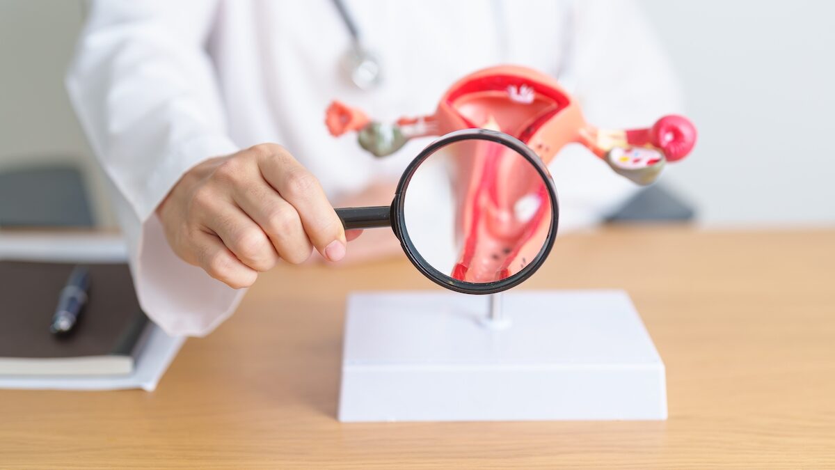
On a medical image, a lesion may appear small. In the body, however, it can be part of a much more active environment than it seems. It is this discrepancy that is shed light on by a study published on October 9, 2026 in Nature Communications : Researchers have mapped endometriosis lesions at very high resolution to understand which cells are next to them, particularly in the case of proximity to glands and nerve structures.
The result does not change the treatment of patients today. Rather, it brings a mechanism track : In some lesions, pain-related nerve cells appear close to immune cells and support cells, a neighborhood that could contribute to inflammation and pain signals.
What does this lesion map reveal?
Endometriosis is characterized by the presence of tissue resembling the endometrium outside the uterus. Its pain remains difficult to explain by a single mechanism, because the disease cannot be reduced to the simple presence of a visible lesion.
“Doctors tend to downplay the symptoms, saying that periods are normally painful and that other women feel the same way.says Caroline Haney, a developmental biologist at Jacksonville University (JAX) and endometriosis patient who led the research. “However, it turns out that the reality is very different. The pain is not just psychological; it is a biological reaction, a real pain signal coming from the body’s neurons.”.
In this work, the team studied eight lesions
coming from the two areas most frequently affected by endometriosis: the ovaries and the peritoneum, the membrane lining the abdominal and pelvic cavities (four superficial peritoneal lesions and four ovarian lesions). The researchers didn’t just seek to identify the cells present. They wanted to know where they were located in relation to each other, as if we were going from a list of inhabitants to a detailed map of a neighborhood.
To do this, they used advanced technologies to reveal which genes are active and the exact position of cells in relation to each other within intact tissue. They therefore give a very detailed reading of the local organization of the lesions, but it does not directly measure the pain felt by a patient.
This nuance matters: the study describes a cellular landscape and possible biological signals. It does not, on its own, predict the intensity of a woman’s symptoms or summarize all the pain related to endometriosis.
Why do nerves, macrophages and fibroblasts matter?
Researchers have discovered that endometriosis lesions are organized in several layers around epithelial glands. Near these glands, three types of cells are concentrated: fibroblasts, which structure tissues and could promote the growth of nerves, macrophages, involved in inflammation, and nociceptive neurons, which transmit pain signals. This organization has been observed in both ovarian and peritoneal lesions.
These observations suggest the existence of a vicious circle between the nervous system and the immune system. Immune cells maintain inflammation, while neurons transmit pain signals. Certain fibroblasts could also promote the development of nerve fibers. These interactions could thus amplify pain and inflammation, contributing to their persistence in certain women with endometriosis.
The further you are from the glands, the less intense these interactions associated with pain are. Other types of sensory neurons are then more present, notably those which detect pressure and movements. These results suggest that pain linked to endometriosis does not only depend on the presence of lesions, but also on the interactions between nerves, immune cells and tissues. This mechanism opens new avenues of research, without yet demonstrating that a treatment targeting these interactions could provide relief to patients.
“Current treatments for endometriosis are not effective for all patients, and even when they are, relief is sometimes only temporary. Our research, which uncovers the neurons involved in injury-related pain, will make it possible to find better solutions for pain management.”declared Caroline Haney.
Is this already a step forward in treating pain?
The authors also used a three-dimensional culture model to study interactions between lesion cells and neurons. This in vitro model makes it possible to observe cellular responses in the laboratory, in particular pro-neuronal signals. By combining these neuronal organoids with human epithelial cells and fibroblasts from patient lesions, the scientists demonstrated that the epithelial cells in combination with the fibroblasts caused an inflammatory and painful state in the neurons.
But this part of the study should not be overinterpreted. A 3D culture is not a clinical trial, and it does not replicate the full experience of chronic pain in a living organism. No new drugs have been tested in female patients in this work.
The main limitation also lies in the size of the sample: eight lesions allow a detailed exploration, not a generalization to all forms of endometriosis. Pain may involve other mechanisms, peripheral or central, which are not resolved by this card alone.
The interest of the study is therefore more fundamental than immediately therapeutic. Today, it allows us to better understand the pathophysiology of this disease in order to be able to tomorrow offer treatments capable of interrupting the inflammatory and painful process induced by the lesions.