In France as elsewhere, women learn that they have a
breast cancer advanced even though their last mammogram was reassuring. These tumors, called “interval cancers”, occur between two examinations and escape traditional screening.
This scenario killed Fatma Caliskanoglu, the aunt of researcher Canan Dagdeviren, despite regular monitoring. From this shock was born the idea of a miniaturized ultrasound system able to monitor the breast much more often, at home or in the office. A team from MIT has just presented a first prototype of this mini 3D ultrasound machine.
Interval cancers: why MIT is banking on portable ultrasound
The works published in the journal Advanced Healthcare Materials remind us of the scale of the problem: interval cancers represent 20 to 30% of cases of breast cancer, with often more aggressive forms. When the tumor is detected at the very early stage, survival approaches 100%; when the diagnosis occurs late, it falls to around 25%.
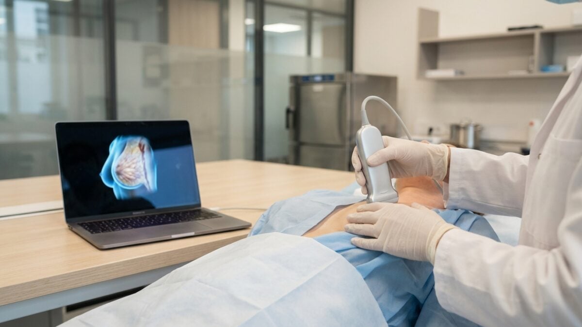
In women at high risk or with dense breasts, more frequent ultrasounds, in addition to mammograms, could identify these tumors earlier between two appointments. Current devices remain bulky, expensive and reserved for expert operators. “Ultrasound imaging has long been confined to hospitals” says Md Osman Goni Nayeem, former postdoctoral fellow at MIT. “To extend its use beyond the hospital environment, we redesigned the complete architecture and introduced a new manufacturing process to make this technology both scalable and practical“.
Conversely, the new device combines a small ultrasound probe, a little smaller than a deck of cards, and a motherboard as an acquisition module, slightly larger than a smartphone. Inside, an ultrasound matrix arranged in a hollow square (CODA architecture) produces wide-angle 3D images while using only 128 elements, compared to 1,024 for a conventional matrix. To view images, the motherboard can be connected to a laptop, making the system fully portable. The module, made from common components, costs around $300 (around €280). And it all works with a simple 5 V power supply!
A pocket 3D probe to monitor the entire breast in just a few gestures
By creating ultrasound systems that are portable and easier to use, the MIT team hopes to make frequent ultrasound exams accessible to more people. “Everything is more compact, which can make it easier to use in rural areas or for people who may have barriers to this type of technology“, explains Professor Canan Dagdeviren, associate professor at MIT. With this system, she explains, more tumors could potentially be detected earlier, which would increase the chances of successful treatment.
 © MIT – Advanced Healthcare Materials
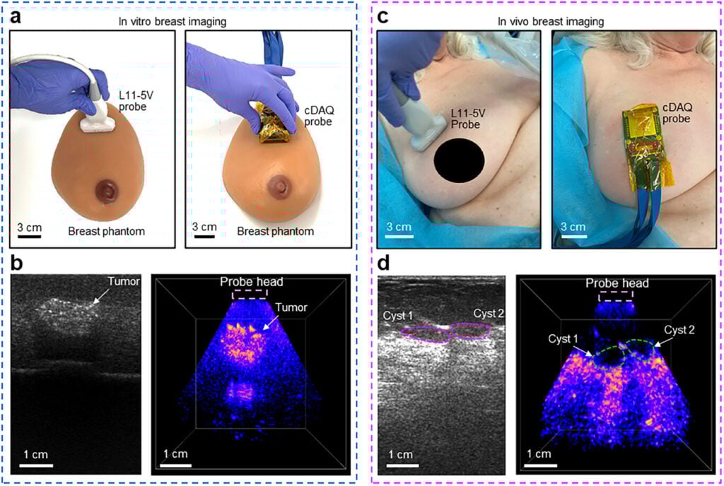
In vitro study on a breast phantom and in vivo study on breast tissue
The sensor was first evaluated on breast phantoms, then on a 71-year-old patient with cysts. The system reconstructed a complete 3D volume of the breast from just two or three probe positions, up to 15 cm deep, with no missing areas. “With our technology, you simply place it gently on the tissue and it can visualize the cysts in their original location and with their original sizes“, describes Canan Dagdeviren.
A larger clinical trial is underway at MIT and Massachusetts General Hospital, ahead of a possible smartphone-connected version.
Towards earlier detection by connecting an AI, but still in the testing phase
Scientists are working on an even smaller version of the data processing system, the size of a fingernail. Their goal is to connect it to a smartphone to view images, which would reduce the size of the device and make it easier to use. They also plan to create a mobile application which, thanks to an artificial intelligence algorithm, would direct the user to the best location to position the ultrasound probe.
Although the current version of the device is easily adaptable for use in a doctor’s office, researchers hope that eventually a more compact version can be integrated into a wearable sensor, allowing use at home for people at high risk of breast cancer.
Professor Dagdeviren is currently working on creating a company to commercialize this technology, with the help of grants from MIT. It was already the origin of a first prototype which we reported on in July 2023.