Every year, breast cancer affects more than 2 million women around the world. After treatments such as radiotherapy or chemotherapy, breast-conserving surgery is often preferred for early-stage breast cancer. The aim of this operation is to remove the tumor while preserving healthy tissue as much as possible. However, due to the difficulty in precisely mapping the tumor during the procedure, it may not be completely removed. As a result, 15-20% of patients require repeat surgery due to insufficient tumor margins. To reduce these reoperations, a team led by Rhys Jones from the University of Western Australia and the Royal Melbourne Hospital developed a portable wireless probe. This probe makes it possible to distinguish tumors from healthy tissues based on their mechanical properties.
A new way to “palpate” breast tumors
The probe exploits the fact that tumors are often stiffer than healthy tissue: a difference that can be felt by the surgeon during the operation.
“The technique presented in this article — stereoscopic optical palpation (SOP) — is inspired by clinical palpation, commonly used by surgeons to identify diseased tissue through the sense of touch” said Rhys Jones, quoted by AIP Publishing.
The portable probe developed by the researchers is based on elastography, a technique that combines imaging with mechanical measurements such as elasticity. Under compression, tumors and healthy tissues have different mechanical properties, which the probe can measure. This also providing a visualization of the fabric, users have additional data to differentiate the two types of fabric.
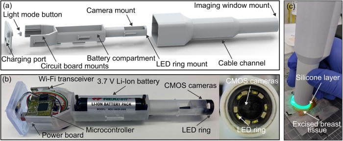
“The wireless probe is an extension of previous tabletop SOP applications. Our approach began by defining the key requirements of the probe, in consultation with surgeons: we opted for an ergonomic and portable format, a field of vision of at least 6 millimeters by 6 millimeters, wireless operation, a minimum battery life of one hour and low cost of materials.” Rhys Jones continued.
The prototype transmits images over Wi-Fi at 15 frames per second and costs around $1,188 for components, or nearly €1,200.
Stiffness maps to guide breast surgery
Performance was evaluated first on a silicone phantom containing a harder inclusion, then on four fresh samples of human breast tissue. Through digital processing, the quality of spatial resolution has been doubled, from 1,034 to 512 micrometers, with an increased stress contrast of approximately 55%.
 © APL Bioengineering
By comparing the mechanical stress maps to histology sections, the team observed that regions of high stress corresponded to tumors, while fat, stroma and canals appeared more “soft”.
Towards fewer re-operations for patients
For patients, better mapping of operating room margins could avoid some of the re-interventions. “Beyond breast-conserving surgery, we believe that the probe could be used in many clinical scenarios where palpation is currently practiced, particularly for the evaluation of skin lesions.“, estimated Rhys Jones in the AIP Publishing publication. He also mentions other potential applications when the cancer is surrounded by soft tissues (prostate, head and neck, etc.).
The authors specify that the device has so far only been tested on excised tissue and that in vivo tests will be necessary before routine use in breast surgery.