What if a simple loss of hearing already left a visible imprint in the brain? For several years, studies have shown that a
age-related hearing loss markedly increases the risk of memory problems and, later, dementia. But the missing link, this precise neuronal mechanism which links ear and cognition, has until now remained very difficult to grasp.
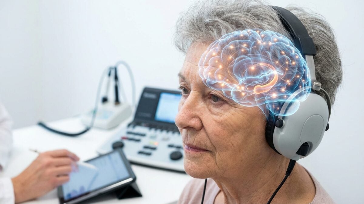
A team led by Ning Li at Tiangong University and Shandong Provincial Hospital followed 55 people with presbycusis and 55 subjects without hearing loss, aged 50 to 74. Thanks to MRI, these researchers believe they have identified a real neurobiological link, which measures how certain brain areas become disorganized when hearing deteriorates.
Hearing loss and dementia: a common association that questions the brain
Presbycusis (age-related hearing loss) mainly affects high-pitched sounds, such as certain consonants, which complicates conversations in noisy environments. We already know that affected people more often present with
cognitive declinewith difficulties with attention, memory or planning. The explanations put forward speak of mental overload to understand speech, sensory deprivation or even social isolation, all of which can weaken the aging brain.
These avenues, however, remained quite indirect: they described possible consequences, without showing where and how the neurons transform. The study published in the journal eNeuro changes scale by focusing on the brain networks themselves, not just behavior. She considers presbycusis to be a neurodegenerative disorder where the ear and the brain age together.
FSR: when brain structure and activity disintegrate together
Concretely, the researchers measured the intensity of the spontaneous activity of a region and its volume of gray matter (schematically the relationship between the structure of the brain and its function) then calculated the function-structure ratio (RFS) by dividing one by the other. In people with presbycusis, they observed an abnormal coupling between atrophy and decreased activity in four key areas linked to sound, memory and decisions: the putamen and the fusiform gyrus (involved in the processing of sounds and speech) as well as the precuneus and the medial superior frontal gyrus (involved in memory and decision-making).
The poorer the hearing and speech recognition thresholds, the more RFS fell in these regions. This decline went hand in hand with poorer results on standardized cognitive tests. The authors summarize: “Decreased RFS correlates with both worsening hearing thresholds and cognitive deterioration“. This ratio thus becomes a true indicator of shared neuronal reorganization:
- It signals brain tissue that is atrophying;
- It reflects local activity which is weakening;
- It indicates less effective integration into networks.
A future biomarker to identify hearing-related dementia risk?
For Ning Li, “The most important thing to remember is that preserving hearing health can protect brain integrity“, he explains, quoted by News-Medical. He believes that variations in the FSR, visible on brain images, could help identify people most at risk of future dementia. External experts already see it as a possible biomarker to detect cognitive impairment linked to hearing loss earlier.