And if the next big breakthrough against
melanoma was contained in an algorithm that sees what the eye does not always perceive.
A new kind of artificial intelligence model
This skin cancer sometimes looks like a harmless mole, making it difficult to diagnose in the office. In 2023, 17,922 cases of cutaneous melanomas were diagnosed in France, according to the Panorama of cancers in France edition 2025. At the same time, the French Society of Dermatology lists 2,928 active dermatologists, after the loss of more than 1,000 specialists in ten years, a context which reinforces the interest in screening tools.
A team from Incheon National University in South Korea, with partners in the United Kingdom and Canada, announces an artificial intelligence model capable of detecting melanoma, which doesn’t just look at the image. The key lies in the way he learns.
An approach that mimics the reasoning of the dermatologist
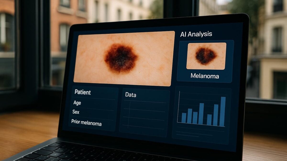
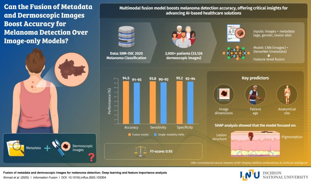
Rather than an image algorithm alone, the team designed a model that merges two sources of information: dermoscopic photos of skin lesions and simple clinical data from the patient, such as age, gender and the anatomical site of the lesion. Clearly, AI is closer to clinical reasoning, where appearance is just one element among others. Trained on the large SIIM ISIC dataset that combines more than 33,000 images with clinical metadata, the system learns subtle connections between what appears on the skin and who the patient is.
 © Incheon National University
Artificial Intelligence Sees What Doctors Can’t: Combining Patient Data and Images to Detect Melanoma
“Skin cancer, particularly melanoma, is a disease for which early detection is absolutely decisive for the chances of survival.” said Professor Gwangill Jeon. “Since melanoma is difficult to diagnose based on visual features alone, I recognized the need for AI convergence technologies that can take into account both imaging data and patient information“.
Melanoma detection with 94.5% accuracy
On the SIIM ISIC test set, the model achieves an accuracy of 94.5% and an F1 score of 0.94, outperforming image-only architectures like ResNet 50 and EfficientNet. The researchers also carried out an analysis of the importance of the variables, showing that the size of the lesion, the age of the patient and the anatomical site weigh heavily in the decision, a step towards a more transparent AI and easier to explain to clinicians.
“The model is not only designed for academic purposes“, emphasizes Gwangill Jeon. “It could be used as a practical tool that could transform real-life melanoma screening. This research can be directly applied to the development of an AI system that analyzes both skin lesion images and basic patient information to enable early detection of melanoma“.
Towards easier screening?
Before widespread adoption by dermatologists, the team points out that additional work is necessary. Professor Jeon explains: “This study represents a step forward toward personalized diagnosis and preventative medicine through the convergence of artificial intelligence technologies.”
The study highlights how multimodal AI can bridge the gap between machine learning and clinical decision-making, paving the way for more accurate, accessible and reliable skin cancer diagnoses.
Ultimately, the model could power skin diagnostic applications on smartphones, telemedicine solutions or office assistance tools. A prospect that could be of interest to France where access to specialists is under pressure.